43 cell wall diagram with labels
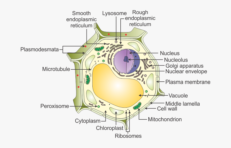
A Labeled Diagram of the Plant Cell and Functions of its Organelles A Labeled Diagram of the Plant Cell and Functions of its Organelles. We are aware that all life stems from a single cell, and that the cell is the most basic unit of all living organisms. ... Unique to plant cells, the cell wall is a fairly rigid, protective wall that resists the strain of physical forces. The cell wall is mainly made up of ... PDF Plant Anatomy: Images and diagrams to explain concepts A diagram of a prokaryotic cell. It lacks organelles and is much smaller and simpler. (LadyofHats Mariana Ruiz. Public Domain). PLANT ANATOMY AND PHYSIOLOGY: IMAGES AND DIAGRAMS TO EXPLAIN 7. 1.2 CELL WALL The cell wall is initially deposited on the surface of the middle lamella. This primary cell wall occurs on the surface of all plant cells ...
03 Label the Cell Diagram | Quizlet Start studying 03 Label the Cell. Learn vocabulary, terms, and more with flashcards, games, and other study tools.
Cell wall diagram with labels
Animal Cell Diagram with Label and Explanation: Cell ... - Collegedunia Diagram of Animal Cell Below is the diagram of the animal cell which shows the organelles present in it. The cell is covered with cytoplasm which consists of cell organelles in it. The nucleus is covered with a rough Endoplasmic Reticulum and other organelles each designed for a specific purpose. Cell wall structure with plant cellular parts description outline diagram Royalty-Free Vector Cell wall structure with plant cellular parts description outline diagram. Labeled educational model components description with hemicellulose, pectin and cellulose microfibril vector illustration. cell wall, plant cell wall structure, vector illustration, components description, vector, wall, educational, diagram, cell, Label Cell Parts | Plant & Animal Cell Activity | StoryboardThat Click "Start Assignment". Find diagrams of a plant and an animal cell in the Science tab. Using arrows and Textables, label each part of the cell and describe its function. Color the text boxes to group them into organelles found in only animal cells, organelles found in only plant cells, and organelles found in both cell types.
Cell wall diagram with labels. Plant Cell: Diagram, Types and Functions - Embibe Exams Plant Cell Wall It is a rigid layer that is composed of cellulose, glycoproteins, lignin, pectin and hemicellulose. It is located outside the cell membrane and is completely permeable. The primary function of a plant cell wall is to protect the cell against mechanical stress and to provide a definite form and structure to the cell. Bacteria in Microbiology - shapes, structure and diagram The bacteria shapes, structure, and labeled diagrams are discussed below. Sizes The sizes of bacteria cells that can infect human beings range from 0.1 to 10 micrometers. Some larger types of bacteria such as the rickettsias, mycoplasmas, and chlamydias have similar sizes as the largest types of viruses, the poxviruses. Structure of Bacterial Cell (With Diagram) - Biology Discussion These are long filamentous, cytoplasmic appendages, 12-30 μm in length, protruding through the cell wall and contain contractile protein flagellin. These are organs of locomotion. Fimbriae or pili: These are thin, short filaments (0.1-1.5 μm x 4 to 8 nm) extruding from the cytoplasmic membrane, also called pili. They are made of protein (pilin). Cell Organelles- Definition, Structure, Functions, Diagram In a plant cell, the cell wall is made up of cellulose, hemicellulose, and proteins while in a fungal cell, it is composed of chitin. A cell wall is multilayered with a middle lamina, a primary cell wall, and a secondary cell wall. The middle lamina contains polysaccharides that provide adhesion and allow binding of the cells to one another.
Structure of Cell Wall (With Diagram) - Biology Discussion Cells with secondary wall consist of five layers a three layered secondary wall, the primary wall and the middle lamella. In some cells, such as primary xylem, the secondary thickening materials are laid down in such a way that various patterns are formed on the cell wall, e.g. annular, spiral, reticulate, scalariform and pitted. Plant Cell Wall Stock Illustrations - 21,398 Plant Cell Wall Stock ... Download 21,398 Plant Cell Wall Stock Illustrations, Vectors & Clipart for FREE or amazingly low rates! New users enjoy 60% OFF. 190,553,602 stock photos online. ... Animal vs plant cell structure comparison with differences outline diagram. Labeled educational inner anatomy description with membrane, cytoplasm and. Coloring page. Plant cell ... Human Cell Diagram, Parts, Pictures, Structure and Functions Diagram of the human cell illustrating the different parts of the cell. Cell Membrane. The cell membrane is the outer coating of the cell and contains the cytoplasm, substances within it and the organelle. It is a double-layered membrane composed of proteins and lipids. The lipid molecules on the outer and inner part (lipid bilayer) allow it to ... Animal Cells: Labelled Diagram, Definitions, and Structure Cell Organelles Plant Cells: Animal Cells: Cell wall: Present (made up of cellulose) Absent: Shape: Rectangular (fixed shape) Round (irregular shape) Vacuole: One, large central vacuole taking up to 90% of cell volume. One or more small vacuoles (much smaller than plant cells). Centrioles: Only present in lower plant forms (e.g. chlamydomonas)
Plant Cells: Labelled Diagram, Definitions, and Structure The cell wall is made of cellulose and lignin, which are strong and tough compounds. Plant Cells Labelled Plastids and Chloroplasts Plants make their own food through photosynthesis. Plant cells have plastids, which animal cells don't. Plastids are organelles used to make and store needed compounds. Chloroplasts are the most important of plastids. PDF Plant Cell Diagram - Edrawsoft Plant Cell Golgi vesicles Golgi apparatus Ribosome Smooth ER(no ribosomes) Nucleolus Nucleus Rough ER(endoplasmic reticulum) Large central vacuole Amyloplast(star ch grain) Cell wall Cell membrane Chloroplast Vacuole membrane Raphide crystal Mitochondrion Druse crystal Label the cell - Teaching resources - Wordwall Label Plant and Animal Cell Labelled diagram by Eawilson the cell Match up by Elenagp9149 5.6 Label the sentence Labelled diagram by Christianjolene Label the Electromagnetic Spectrum Labelled diagram by Elizabetheck G6 G7 G8 Science 5.7 Label the sentence Labelled diagram by Christianjolene The cell Anagram by Thepowerhouse G7 G8 Science Plant Cell Diagram | Science Trends A plant cell diagram, like the one above, shows each part of the plant cell including the chloroplast, cell wall, plasma membrane, nucleus, mitochondria, ribosomes, etc. A plant cell diagram is a great way to learn the different components of the cell for your upcoming exam. Plants are able to do something animals can't: photosynthesize.
A Draw A Neat Diagram Of A Plant Cell And Label The - Diagram Of Plant Cell With Labelling ...
Animal Cell Labelling Activity | Primary Resources | Twinkl A colorful resource which covers the parts of an animal cell including the nucleus, cell wall, cytoplasm, and mitochondria. Lower, middle and higher ability versions are available. ... this Plant Cell Diagram is a similar labelling activity for plant cells. ... you could try using this Polar Bear Animal Diagram with Labels.
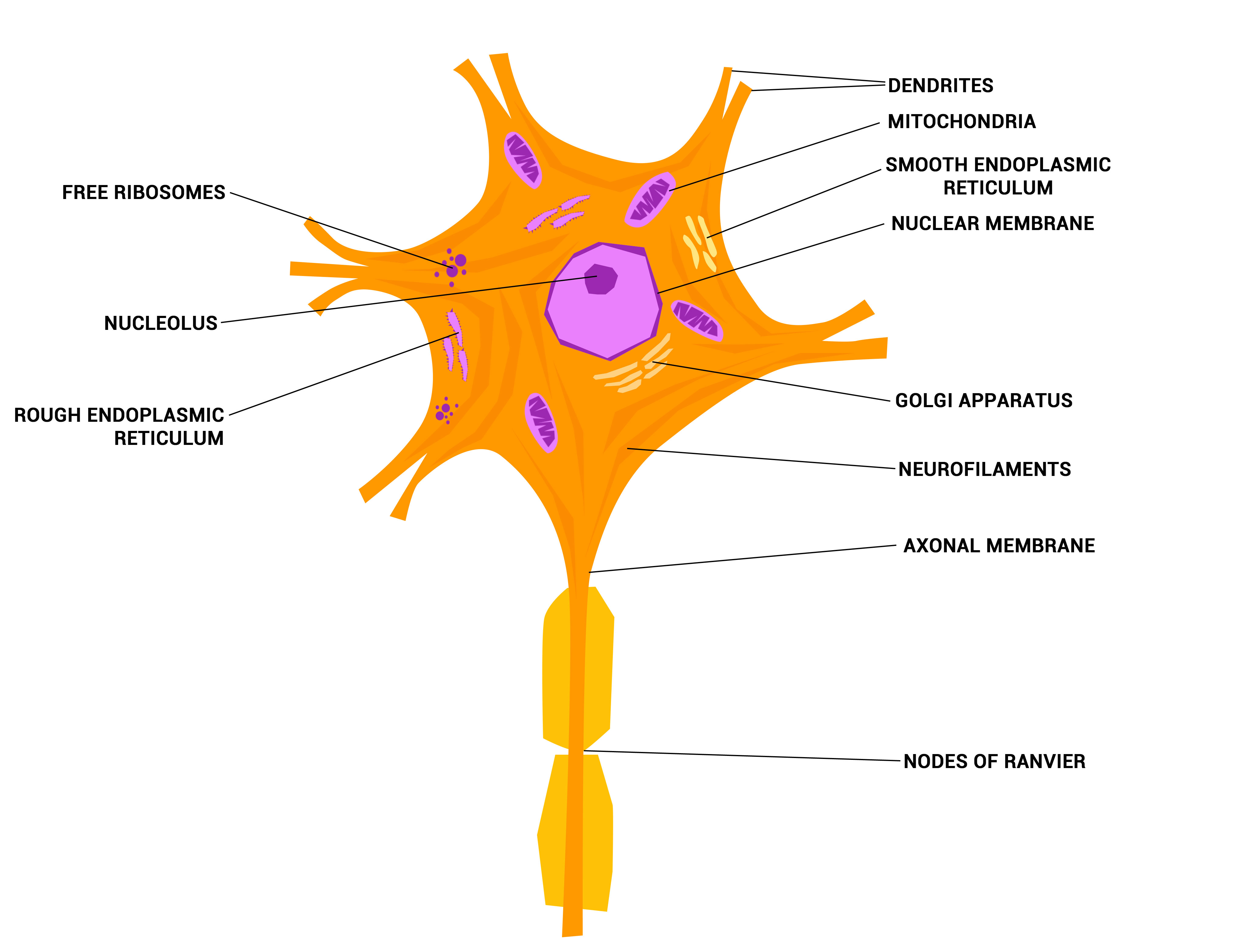
Neurons - The crazy wires in our body. | Doc Jana
Elodea Leaf Cell Diagram Elodea Leaf Cell Diagram The Elodea leaf is composed of two layers of cells. Only one layer of cells is in focus when using the high. Examining elodea (pondweed) under a compound microscope. solution) and a coverslip and observe the chloroplasts (green structures) and the cell walls.
Post a Comment for "43 cell wall diagram with labels"