44 picture of compound microscope with labels and functions
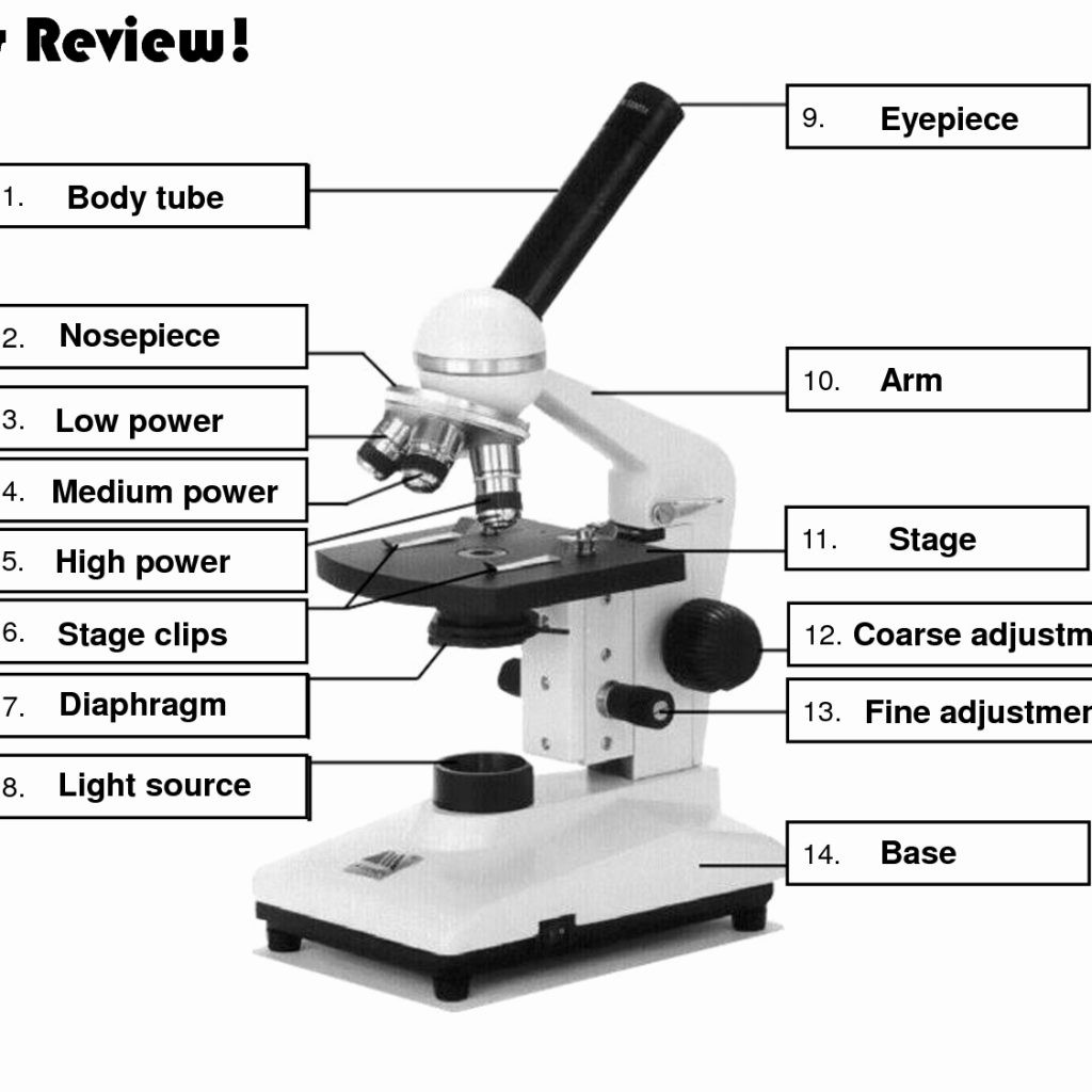
Parts of the Microscope with Labeling (also Free Printouts) Let us take a look at the different parts of microscopes and their respective functions. 1. Eyepiece it is the topmost part of the microscope. Through the eyepiece, you can visualize the object being studied. Its magnification capacity ranges between 10 and 15 times. 2. Body tube/Head It is the structure that connects the eyepiece to the lenses. Label Parts Of A Compound Microscope Teaching Resources | TpT This presentation contains: high quality images, 3 formative assessments and a virtual microscopic slide show. Main concepts: History of the discovery of the microscope Microscope parts and functions label the parts of a microscope 3 formative assessments how to use, carry and store the microscope how to calculate magnificati
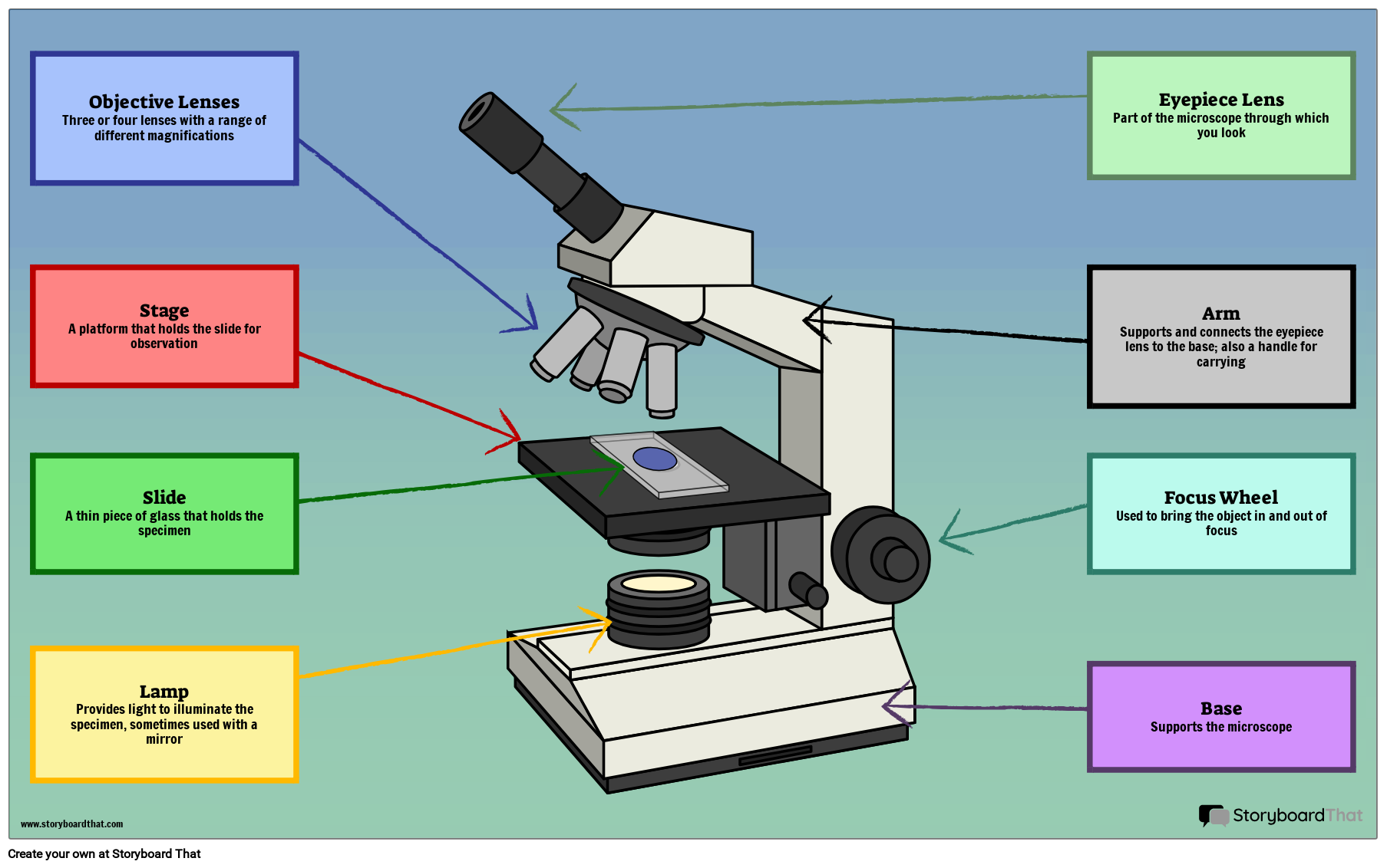
Compound Microscope Parts, Functions, and Labeled Diagram Compound Microscope Definitions for Labels. Eyepiece (ocular lens) with or without Pointer: The part that is looked through at the top of the compound microscope. Eyepieces typically have a magnification between 5x & 30x. Monocular or Binocular Head: Structural support that holds & connects the eyepieces to the objective lenses.

Picture of compound microscope with labels and functions
Diagram of a Compound Microscope - Biology Discussion 1. It is noted first that which objective lens is in use on the microscope. 2. Stage micrometer is positioned in such a way that it is in the field of view. 3. The eyepiece is rotated so that the two scales, the eyepiece or ocular scale and the stage micrometer scale, are parallel. 4. What is a Compound Microscope? - New York Microscope Company A compound microscope is an instrument that is used to view magnified images of small specimens on a glass slide. It can achieve higher levels of magnification than stereo or other low power microscopes and reduce chromatic aberration. It achieves this through the use of two or more lenses in the objective and the eyepiece. Parts of a Compound Microscope and Their Functions Compound microscope magnification is determined by multiplying the eyepiece and objective powers. When viewed through a 5X eyepiece with a 10X objective, an item is magnified 5 x 10=50 times. The magnification is 10 x 45 = 450 times when using a 10X eyepiece and a 45X objective. How to Use the Compound Microscope
Picture of compound microscope with labels and functions. Timeline of United States inventions (1946–1991) - Wikipedia A Scanning Acoustic Microscope (SAM) is a device which uses focused sound to investigate, measure, or image an object. It is commonly used in failure analysis and non-destructive evaluation. The first scanning acoustic microscope was co-invented in 1974 by C. F. Lemons and R. A. Quate at the Microwave Laboratory of Stanford University. Compound Microscope Parts, Function, & Diagram | What is a Compound ... It is the piece of the compound light microscope that a person looks through to view the image. It is located at the top of the body tube. The eyepiece contains a lens that also helps to enlarge... Compound Microscope Parts - Labeled Diagram and their Functions - Rs ... Basically, compound microscopes generate magnified images through an aligned pair of the objective lens and the ocular lens. In contrast, "simple microscopes" have only one convex lens and function more like glass magnifiers. [In this figure] Two "antique" microscopes played significant roles in the history of biology. Labeling the Parts of the Microscope | Microscope World Resources Labeling the Parts of the Microscope. This activity has been designed for use in homes and schools. Each microscope layout (both blank and the version with answers) are available as PDF downloads. You can view a more in-depth review of each part of the microscope here.
Microscope picture label Flashcards | Quizlet Start studying Microscope picture label. Learn vocabulary, terms, and more with flashcards, games, and other study tools. ... Microscope Parts and Functions. 32 terms. zukowskie. Science Microsope. 27 terms. 15jprice. The Compound Light Microscope Parts. 14 terms. mtps_anna. Other sets by this creator. 18-19 YEAR IN REVIEW. 479 terms. kfire ... Compound Microscope - Types, Parts, Diagram, Functions and Uses Compound microscope - It is an optical instrument consists of two convex lenses of short focal lengths primarily used for observing a highly magnified image of minute objects. Lenses Simple microscope - It has a convex lens. It uses only one lens to magnify objects. An example of a simple microscope is a magnifying glass. Microscope Parts and Functions First, the purpose of a microscope is to magnify a small object or to magnify the fine details of a larger object in order to examine minute specimens that cannot be seen by the naked eye. Here are the important compound microscope parts... Eyepiece: The lens the viewer looks through to see the specimen. Parts of a Compound Microscope (And their Functions) List of Microscope Parts and their Functions 1. Ocular Tubes (Monocular, Binocular & Trinocular) The ocular tubes, are to tubes that lead from the head of the microscope out to your eyes. On the end of the ocular tubes are usually interchangeable eyepieces (commonly 10X and 20X) that increase magnification.
Compound Microscope Labeled Diagram - Quizlet QUESTION. The total magnification of a specimen being viewed with a 10X ocular lens and a 40X objective lens is. 15 answers. QUESTION. a mosquito beats its wings up and down 600 times per second, which you hear as a very annoying 600 Hz sound. if the air outside is 20 C, how far would a sound wave travel between wing beats. 2 answers. Compound microscope - their parts and function - Microscopy4kids The term "compound" refers to the microscope having more than one lens. Compound microscopes generate magnified images through an aligned pair of the objective lens and the ocular lens. In contrast, "simple microscopes" have only one convex lens and function more like glass magnifiers. Eyepiece (ocular lens) Biology II Mid-term Study Guide Flashcards | Quizlet Labels may be used more than once. Bacteria:include photosynthetic and nitrogen-fixing prokaryotes; unicellular; include e coli, vibrio cholera, and salonella typhoides; contain no membrane bound organelles; contain plasma membranes made of phospholipids and cell walls made of peplidoglycan Compound Microscope - Diagram (Parts labelled), Principle and Uses Compound Microscope - Diagram (Parts labelled), Principle and Uses As the name suggests, a compound microscope uses a combination of lenses coupled with an artificial light source to magnify an object at various zoom levels to study the object. A compound microscope: Is used to view samples that are not visible to the naked eye

Microscopes Online Worksheet Answers - Nidecmege
Microscope Types (with labeled diagrams) and Functions A compound microscope: Is used to view samples that are not visible to the naked eye Uses two types of lenses - Objective and ocular lenses Has a higher level of magnification - Typically up to 2000x Is used in hospitals and forensic labs by scientists, biologists and researchers to study micro organisms Compound microscope labeled diagram

Compound Microscope Diagram Easy To Draw - Micropedia
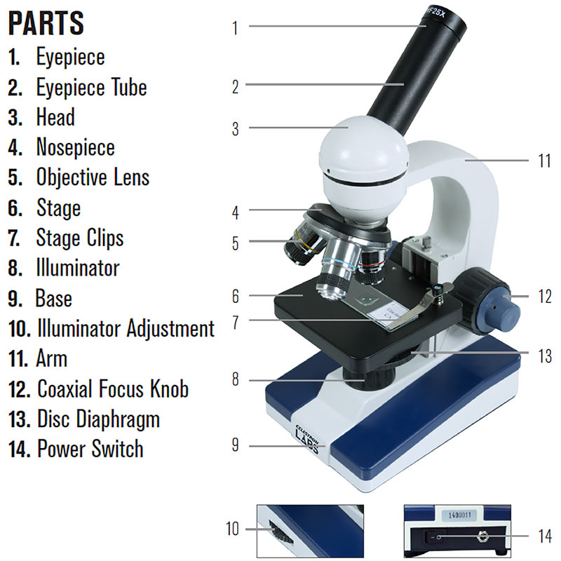
Microscope Parts, Function, & Labeled Diagram - slidingmotion Condenser. The condenser is to focus the light, which passes from the microscopic illuminator to the specimen. This condenser is located just below the diaphragm. This diaphragm is one of the important parts of the compound microscope which will help to get an accurate and sharp image. The condenser has a magnification power of 400X and above.
31 Picture Of Microscope With Label - Labels Database 2020
Targeting LIPA independent of its lipase activity is a ... Jun 02, 2022 · Raj and colleagues show that ERX-41 inhibits lipase-independent functions of LIPA and induces ER stress in different tumor types, providing a therapeutic strategy in PDX models of pancreas ...

Anatomy and Physiology I Coursework: Microscope A+P
Parts of a microscope with functions and labeled diagram Optical parts of a microscope and their functions The optical parts of the microscope are used to view, magnify, and produce an image from a specimen placed on a slide. These parts include: Eyepiece - also known as the ocular. This is the part used to look through the microscope. Its found at the top of the microscope.

Compound Light Microscope Labeled - Made By Creative Label
Label the microscope - Science Learning Hub All microscopes share features in common. In this interactive, you can label the different parts of a microscope. Use this with the Microscope parts activity to help students identify and label the main parts of a microscope and then describe their functions. Drag and drop the text labels onto the microscope diagram.
Binocular Microscope Sketch at PaintingValley.com | Explore collection of Binocular Microscope ...
A Study of the Microscope and its Functions With a Labeled Diagram A Study of the Microscope and its Functions With a Labeled Diagram To better understand the structure and function of a microscope, we need to take a look at the labeled microscope diagrams of the compound and electron microscope. These diagrams clearly explain the functioning of the microscopes along with their respective parts. M mooketsi

Microscope Parts and Functions
Parts of a Compound Microscope - Labeled (with diagrams) A compound microscope is known as a high-power microscope that enables you to achieve a high level of magnification. Smaller specimens can be thoroughly viewed using a compound microscope. Let us take a look at the different parts of a compound microscope and understand each key component.
Celestron Labs Compound Microscope CM1000C
(PDF) Taiz & Zeiger- Plant Physiology | Munish K Bansal ... Enter the email address you signed up with and we'll email you a reset link.
Post a Comment for "44 picture of compound microscope with labels and functions"