39 microscope diagram with labels and definitions
Microscope Parts & Functions - AmScope Iris Diaphragm: Found on high power microscopes under the stage, the diaphragm is, typically, a five hole-disc with each hole having a different diameter. It is used to vary the light that passes through the stage opening and helps to adjust both the contrast and resolution of a specimen. It is particularly useful at higher powers. Microscope Types (with labeled diagrams) and Functions A compound microscope: Is used to view samples that are not visible to the naked eye. Uses two types of lenses - Objective and ocular lenses. Has a higher level of magnification - Typically up to 2000x. Is used in hospitals and forensic labs by scientists, biologists and researchers to study micro organisms. Compound microscope labeled diagram.
Parts of a Microscope and their function - Microbiology Note The Aperture: It is a hole on the microscope stage. The light transmitted through this hole from the source to the specimen. Microscopic illuminator: This is basically a light source for a microscope, found at the base. It is a constant light source (110 volts in the US) that lights up through the slide.

Microscope diagram with labels and definitions
PDF The Microscope Parts and Use - Plainview the year 1590. The compound microscope uses lenses and light to enlarge the image and is also called an optical or light microscope (vs./ an electron microscope) . The simplest optical microscope is the magnifying glass and is good to about ten times (10X) magnification. The Compound Microscope- Definition, Labeled Diagram, Principle, Parts, Uses A standard Microscope has three to four Objective Lenses which range from 4X to 100X. Stage Clips are metal clips that held the slide in place. Arm and Base. The Arm connects the Body Tube to the base of the Microscope. The Base supports the Microscope and its where Illuminator. Illuminator and Stage. The illuminator is the light source for a microscope. Microscope Glossary of Terms: Microscope A-Z - Microscope and ... The purpose is to allow 3-dimensional movement with the microscope usually for studying large subjects such as textile work. B . Barrel Focus - When the stage is fixed and the body tube of the microscope is moved to focus the objective aperture. Base - The bottom of the microscope on which the body attaches to. The base will have a stand and may have a clamp for securing a microscope to a desk.
Microscope diagram with labels and definitions. Types of Microscopes: Definition, Working Principle, Diagram ... Types of Microscopes: Definition, Working Principle, Diagram, Applications, FAQs There are 5 types of microscopes. These microscope types find applications in different fields. Simple microscope, compound microscope, stereo microscope, scanning probe microscope, electron microscope are explained. Microscope Label Interactive Worksheets & Teaching Resources | TpT 12. $1.89. PDF. Students will complete a timeline of the history of the microscope, label a diagram, and create a pocket foldable with terms and definition cards. The timeline can be completed according to the teacher's directions or like the answer key example. Optional cut & paste images and a QR code are a. Simple Microscope - Parts, Functions, Diagram and Labelling Simple Microscope - Parts, Functions, Diagram and Labelling A microscope is one of the commonly used equipment in a laboratory setting. A microscope is an optical instrument used to magnify an image of a tiny object; objects that are not visible to the human eyes. Table of Contents The common types of microscopes are: What is a Simple microscope? Microscope Diagram With Definitions - 14 best images of microscope ... Here are a number of highest rated Microscope Diagram With Definitions pictures upon internet. We identified it from obedient source. Its submitted by dealing out in the best field. We take this nice of Microscope Diagram With Definitions graphic could possibly be the most trending subject considering we allowance it in google gain or facebook.
Diagram of a Compound Microscope - Biology Discussion Magnification may be defined as the degree of enlargement of the image of an object provided by the microscope. Magnification by a microscope is the product of the individual magnifying ability of the oculars and the objectives. For example if the ocular is 10X, and objective is 40X, the specimen is magnified 400 times. microscope | Types, Parts, History, Diagram, & Facts | Britannica microscope, instrument that produces enlarged images of small objects, allowing the observer an exceedingly close view of minute structures at a scale convenient for examination and analysis. Although optical microscopes are the subject of this article, an image may also be enlarged by many other wave forms, including acoustic, X-ray, or electron beam, and be received by direct or digital ... Label the microscope — Science Learning Hub All microscopes share features in common. In this interactive, you can label the different parts of a microscope. Use this with the Microscope parts activity to help students identify and label the main parts of a microscope and then describe their functions. Drag and drop the text labels onto the microscope diagram. If you want to redo an answer, click on the box and the answer will go back to the top so you can move it to another box. PDF Parts of the Light Microscope - Science Spot B. NOSEPIECE microscope when carried Holds the HIGH- and LOW- power objective LENSES; can be rotated to change MAGNIFICATION. Power = 10 x 4 = 40 Power = 10 x 10 = 100 Power = 10 x 40 = 400 What happens as the power of magnification increases?
Parts of the Microscope with Labeling (also Free Printouts) Parts of the Microscope with Labeling (also Free Printouts) A microscope is one of the invaluable tools in the laboratory setting. It is used to observe things that cannot be seen by the naked eye. Table of Contents 1. Eyepiece 2. Body tube/Head 3. Turret/Nose piece 4. Objective lenses 5. Knobs (fine and coarse) 6. Stage and stage clips 7. Aperture PDF Definitions of the Parts of the Microscope - UAlberta Definitions of the Parts of the Microscope © Heather Kroening May 2001 Arm - The arm of the microscope supports the body tube. Body Tube - The body tube is a hollow tube through which light travels from the objective to the ocular. It contains a prism at the base of the tube that bends the light rays so they can enter the inclined tube. Biology Microscope Labeling and Definitions (Light/Compound Microscope ... Biology Microscope Labeling and Definitions (Light/Compound Microscope) study guide by joshb9051 includes 8 questions covering vocabulary, terms and more. Quizlet flashcards, activities and games help you improve your grades. Compound Microscope Parts - Labeled Diagram and their Functions The eyepiece (or ocular lens) is the lens part at the top of a microscope that the viewer looks through. The standard eyepiece has a magnification of 10x. You may exchange with an optional eyepiece ranging from 5x - 30x. [In this figure] The structure inside an eyepiece. The current design of the eyepiece is no longer a single convex lens.
All Saints Online: Microscope Part Functions
Microscope Diagram With Labels And Functions Label Microscope Diagram Enchantedlearning Com The Functional Parts Of The Microscope Enfo Diagram Quiz On Compound Microscope Parts And Functions The Microscope Teaching Resources Https Bluegrass Kctcs Edu Education Training Media Natural Sciences Biology Bio137 Lab2 137labexercise2 Pdf Parts Of A Microscope ...

32 Microscope Label And Functions - Modern Label Ideas
Compound Microscope Parts, Functions, and Labeled Diagram Compound Microscope Definitions for Labels Eyepiece (ocular lens) with or without Pointer : The part that is looked through at the top of the compound microscope. Eyepieces typically have a magnification between 5x & 30x.

Animal Cell Labeled And Functions Quizlet / Animal Cell Labeling And All Definitions For ...
Compound Microscope: Definition, Diagram, Parts, Uses, Working ... - BYJUS A microscope with a high resolution and uses two sets of lenses providing a 2-dimensional image of the sample. The term compound refers to the usage of more than one lens in the microscope. Also, the compound microscope is one of the types of optical microscopes. The other type of optical microscope is a simple microscope.

Biology 2201
PDF Parts of a Microscope Printables - Homeschool Creations Label the parts of the microscope. You can use the word bank below to fill in the blanks or cut and paste the words at the bottom. Microscope Created by Jolanthe @ HomeschoolCreations.net. Parts of a eyepiece arm stageclips nosepiece focusing knobs illuminator stage objective lenses

Fig. 32.7 A=A clusture of male cones ; B=longitudinal section of a male ... | cones | Pinterest | Ss
Microscope Parts and Functions The specimen is placed on the glass and a cover slip is placed over the specimen. This allows the slide to be easily inserted or removed from the microscope. It also allows the specimen to be labeled, transported, and stored without damage. Stage: The flat platform where the slide is placed.

32 Compound Light Microscope Label - Labels 2021
A Study of the Microscope and its Functions With a Labeled Diagram These labeled microscope diagrams and the functions of its various parts, attempt to simplify the microscope for you. However, as the saying goes, 'practice makes perfect', here is a blank compound microscope diagram and blank electron microscope diagram to label. Download the diagrams and practice labeling the different parts of these fascinating instruments.
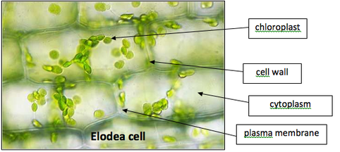
Lab Micrographs - ChristinaLovemicroscopy
16 Parts of a Compound Microscope: Diagrams and Video Body of the Microscope In compound microscopes with two eye pieces there are prisms contained in the body that will also split the beam of light to enable you to view the image through both eye pieces. 2. Arm The arm of the microscope is another structural piece. The arm connects the base of the microscope to the head/body of the microscope.
Post a Comment for "39 microscope diagram with labels and definitions"